
Description
Overview
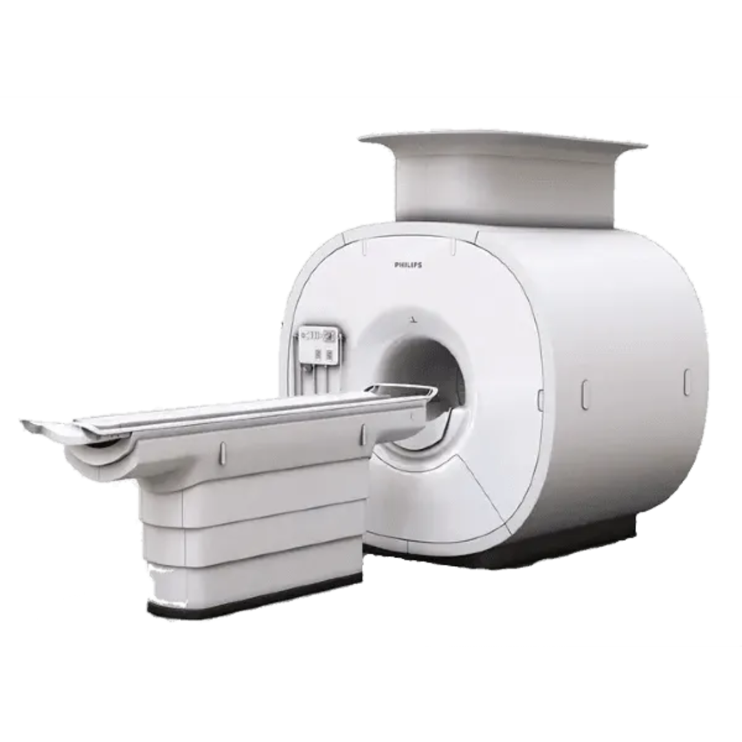
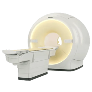
The Philips Multiva 1.5T is built to address growing patient volumes and operational cost pressures by making high-performance imaging routine and repeatable. Designed with an ultra-compact $1.57\text{-meter}$ short-bore magnet, the system offers an inviting entry flare ($119\text{ cm}$) and a $53\text{ cm}$ usable Field of View (FOV) to minimize patient claustrophobia while delivering consistent fat suppression and spatial resolution.
At the core of Multiva’s image performance is oStream Direct Digital Sampling (DDS). By executing analog-to-digital signal conversion right inside the scan room at the magnet frame, oStream eliminates noise pick-up along long cable runs to ensure raw data integrity. Combined with FlexStream workflow design—which drastically reduces the need for frequent coil changes—and SENSE parallel imaging, the Multiva platform allows radiology departments to achieve full diagnostic acquisitions in as little as 10 minutes per exam.
Key Features & Advanced Technologies
-
oStream Direct Digital Receiver Architecture: Digitizes signals directly at the magnet in the scan room, providing high signal clarity, dynamic range up to $162\text{ dB}$, and sub-millisecond $TR/TE$ timing for high-resolution 2D and 3D imaging.
-
FlexStream Workflow Design: Integrated table coils combined with lightweight surface arrays allow technologists to image multiple anatomical zones without swapping coils, cutting total setup and positioning time by up to $40\%$.
-
SmartAssist Automation Platform: AI-driven workflow assistant that automates routine operator tasks, standardizes slice placement via ExamCards, and provides consistent diagnostic outcomes across different staff shifts.
-
SENSE Parallel Imaging: Industry-standard parallel acceleration software that enables up to $8\times\text{–}16\times$ scan acceleration factors, dramatically shortening patient breath-holds and reducing motion artifacts.
-
SofTone Acoustic Reduction Technology: Suppresses acoustic gradient noise levels without compromising gradient performance or altering tissue contrast appearance.
-
mDIXON XD & Advanced Clinical Applications: Provides multi-contrast imaging (water, fat, in-phase, out-of-phase) in a single rapid acquisition, alongside optional non-contrast renal angiography, ASL neuro perfusion, and whole-body oncology staging.
-
Compact Footprint & Low Cost of Ownership: Requires a minimal installation area of just $19.5\text{ m}^2$ ($210\text{ sq ft}$) and features actively shielded zero-boil-off magnet technology with low power consumption.
Technical Specifications
| Parameter | Specification |
| Field Strength | $1.5\text{ Tesla}$ Superconducting |
| Magnet Length | $1.57\text{ m}$ ($5\text{ ft } 2\text{ in}$) Ultra-Compact |
| Bore Opening | $60\text{ cm}$ Inner Bore with $119\text{ cm}$ Horizontal Entry Flare |
| Homogeneous Field of View (FOV) | Up to $53\text{ cm} \times 53\text{ cm} \times 45\text{ cm}$ |
| Gradient Performance | $33\text{ mT/m}$ Peak Amplitude / Up to $208\text{ T/m/s}$ Slew Rate |
| RF Receive Channels | 16-Channel Scalable Direct Digital Receiver (DDS) |
| RF Transmit Power | $18\text{ kW}$ Single-Channel Air-Cooled Transmit Amplifier |
| Parallel Acceleration Engine | SENSE Technology |
| Patient Table Capacity | $250\text{ kg}$ ($550\text{ lbs}$) Wide Patient Support |
| Minimum Siting Area | $19.5\text{ m}^2$ ($210\text{ sq ft}$) Scan/Control/Technical Room Suite |
Reviews
There are no reviews yet.